Your esophageal cancer specialist.
Because I have set myself the goal of ensuring the best possible life and quality of life for our patients suffering from esophageal cancer, I devote a large part of my energy to this topic. All members of our highly trained and experienced interdisciplinary team do a great job. I myself am considered an internationally recognized specialist in esophageal cancer.
Our individual treatment concept for esophageal cancer.
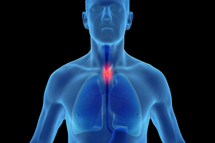
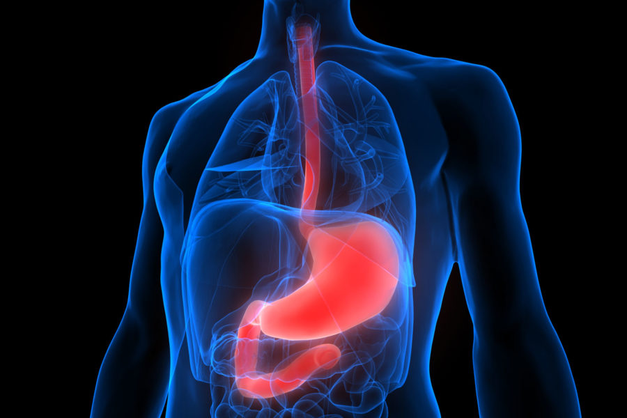
The oesophagus is a 30 - 40 cm long organ that runs through the throat, chest and abdominal cavity region and has a close positional relationship to the large blood vessels aorta, lung and heart. Since the oesophagus and trachea are directly next to each other, the trachea is closed off by the epiglottis for safety when swallowing.
Due to the complex anatomy is the treatment of esophageal diseases only Esophageal specialists and specialty centers The treatment concept should be tailored to the patient's individual needs - life situation, tumor biology and personal wishes. The treatment concept should be individually tailored to the patient's life situation, tumor biology and personal wishes.
Types of oesophageal cancer
In the oesophagus, oesophageal specialists distinguish between three different types of malignant cancer (oesophageal carcinoma).
Squamous cell carcinoma may affect all sections of the oesophagus. It is usually caused by smoking, often in combination with alcohol, but can also be virus-associated and thus also occur in younger people.
The so-called adenocarcinoma (AEG) of the oesophagogastric junction often develops due to tissue changes of the junction between the oesophagus and the stomach triggered by heartburn.
Oesophageal specialists distinguish between:
- AEG I (in the oesophagus)
- AEG II (between oesophagus and stomach)
- and AEG III (at the uppermost part of the stomach).
This tumour is also called a "prosperity tumour" because there may be a connection with a long-standing reflux disease (heartburn).

Oesophageal cancer treatment
If oesophageal cancer is diagnosed, the extent of the tumour determines the respective therapy. If there are no metastases in other organs, surgery is the best solution.
Advanced carcinomas
If the tumour (whether squamous cell carcinoma or adenocarcinoma of the oesophagogastric junction) has crossed the mucosal border, oesophageal specialists choose from the following procedures:
The surgical procedure is based on a concept that is tailored to you and the tumour disease. Gentle, state-of-the-art, minimally invasive and robot-assisted removal of the oesophagus is used.
If the oesophagus has to be partially or completely removed, it can be reconstructed and replaced by various other organs such as the stomach, the small intestine or the colon. This depends on the patient's individual situation and is tailored to you.
Above a certain tumour size, experts achieve the best healing success with a multimodal, interdisciplinary concept. In this case, before and after the operation, additional medical tumour therapy and/or radiotherapy is carried out.
If surgery cannot be performed on patients due to their general condition and the presence of metastases, drug-based tumour therapy and/or radiotherapy are available - supplemented by endoscopictherapy.

Your interdisciplinary treatment team
This type of treatment requires close interaction between a wide range of disciplines, such as oncology, radiology, endoscopy, radiotherapy and surgery. Therefore, it is only possible at centres. We have the necessary practical experience to perform this difficult procedure.
Life after the operation
Generally, no special rehabilitation is necessary after the hospital stay.
We recommend that you have a nutritional consultation with us at the hospital. In principle, the consumption of all foods and drinks in somewhat smaller portions is possible again. Thanks to the latest surgical techniques, a very good quality of life can be achieved even after complete removal of the oesophagus.
After the operation, you will receive an individual aftercare pass from us, in which a regular check of the healing success (tumour aftercare) is carried out.

